
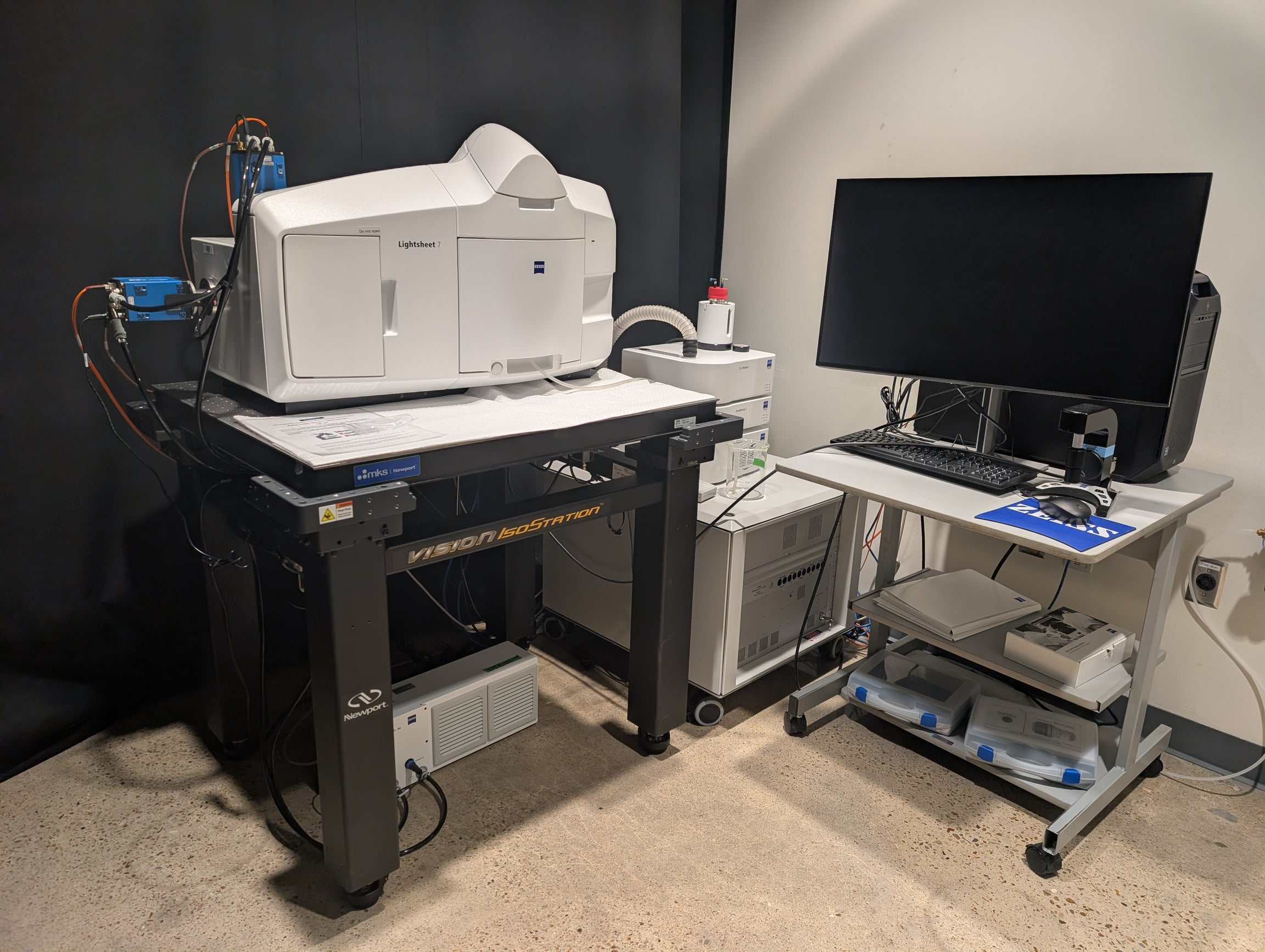
ZEISS Light-Sheet 7
LOCATION
ABL 108
PRICING
Rice User Fee: $25/hr
Non-Profit Fee: $39/hr
For Profit Fee: $125/hr
Rice Training Fee: $50/hr
Non-Profit Training Fee: $78/hr
For Profit Training Fee: $250/hr
CONTACTS
Training Contact:
Subash Godar (Instrument manager) – sg165@rice.edu
Hannah Johnson (Super User) – hlj3@rice.edu
A Budi Utama (Supervisor) – budiutama@rice.edu
Rosa Uribe (PI) – rosa.uribe@rice.edu
DESCRIPTION
The ZEISS Light-sheet 7 is an advanced light sheet fluorescence microscope (LSFM) designed for fast, gentle, and long-term imaging of large, intact specimens. It is particularly well-suited for observing dynamic processes in living organisms, tissues, and cells, as well as for high-resolution imaging of optically cleared samples in their native 3D context, providing a complete picture of cellular and tissue-level events. Its core strength lies in its ability to non-invasively image large specimens with exceptional speed and clarity. The system’s unique illumination method, where a thin plane of light is scanned through the sample, drastically minimizes phototoxicity and photobleaching. This gentle approach makes it the ideal platform for long-term, multi-day experiments on delicate living specimens, such as developing embryos, organoids, or tissues. The performance of the light-sheet 7 is characterized by its remarkable speed and optical precision. The microscope is equipped with a high quantum efficiency sCMOS camera sensor, enabling acquisition speeds of up to 40 fps at full resolution. Additionally, the LSFM uses two separate light paths: a thin sheet of light illuminates a single plane of the sample, while a perpendicular detector captures the image. This approach provides inherent optical sectioning without the need for a pinhole or post-processing. Since the camera captures the entire plane at once, LSFM is much faster and uses significantly less light than confocal or other scanning microscopes. The system’s high-performance objectives provide outstanding sub-cellular resolution. Furthermore, the microscope’s Pivot Scan technology effectively eliminates common shadowing artifacts, while dual-sided illumination ensures uniform light penetration across large, heterogeneous samples, resulting in high-quality, artifact-free image. Moreover, the LSFM offers a highly adaptable and user-friendly experience. Its modular design allows it to be customized for a wide range of applications, from imaging delicate live samples in an environmentally controlled chamber to visualizing cleared tissue volumes up to 2 cm in size. The system’s compatibility with diverse optical clearing protocols and its support for a broad range of refractive indices (1.33-1.58) make it a versatile tool for researchers across different disciplines. The LSFM’s Multiview capability provides a powerful solution for overcoming imaging artifacts in large or dense samples. It works by automatically rotating the specimen and acquiring multiple 3D datasets from different angles. The intuitive ZEN software simplifies complex acquisition workflows, enabling multi-view imaging and tiling of large samples, all while providing integrated solutions for data processing and analysis. FEATURES 1) Microscope 2) Detection optics 3) Illumination optics 4) Lasers 5) Sample Chambers 6) Cameras 7) Software