
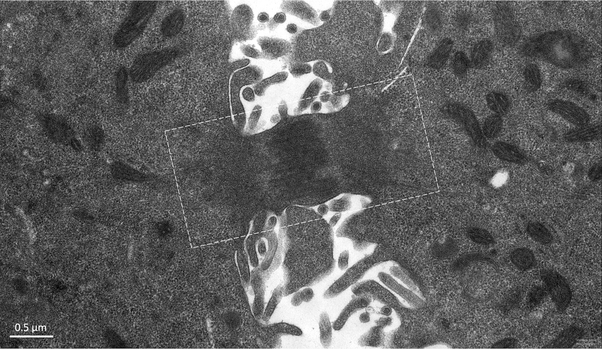
Transmission electron microscopy (TEM) image of a macrophage cell undergoing mitosis (late telophase), with microtubules of the spindle apparatus boxed.
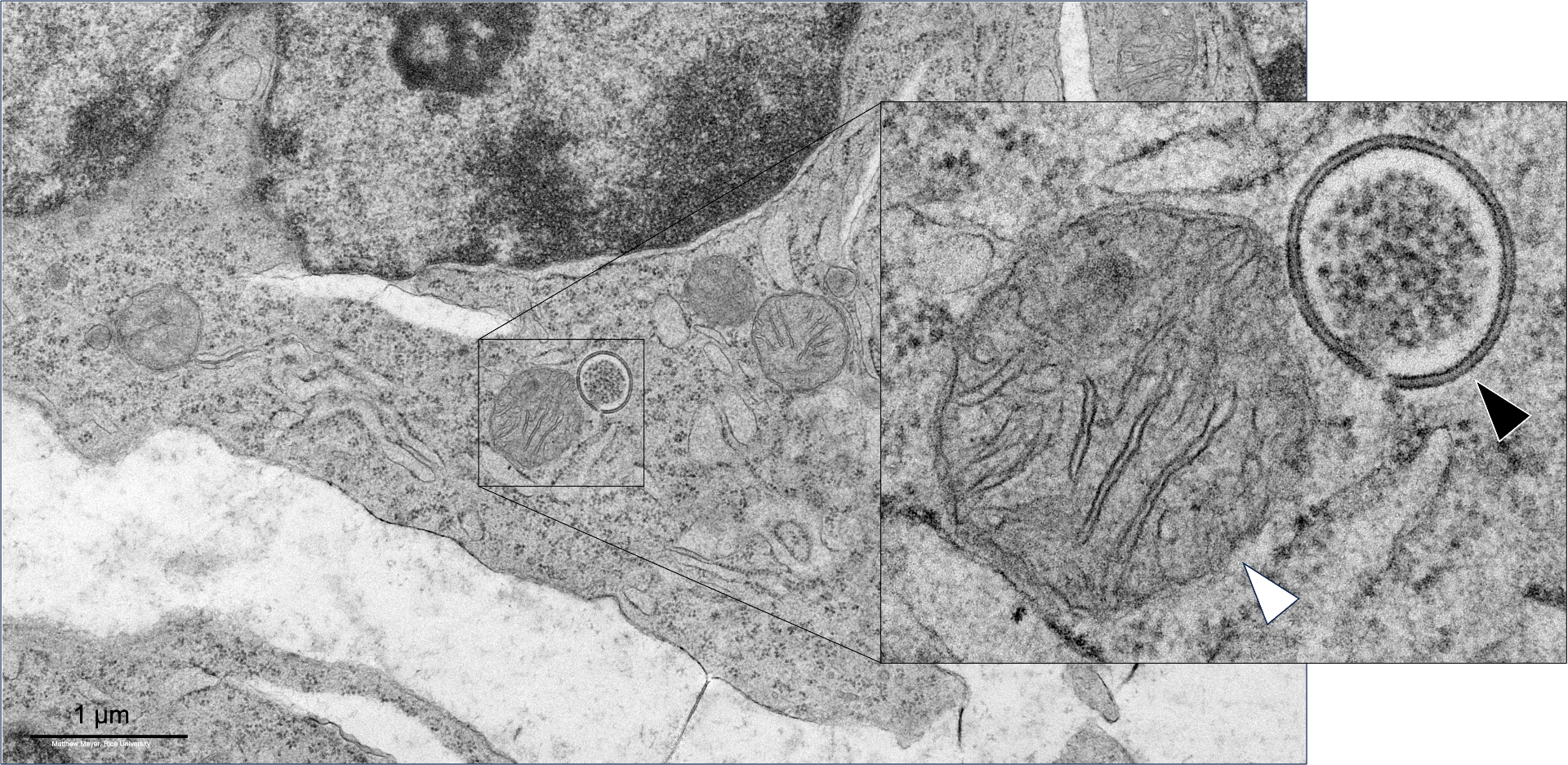
Transmission electron microscopy (TEM) image of a mitochondria in contact with a lysosome, located in an ovarian follicular cell. Mitochondria (white arrow), lysosome (black arrow).
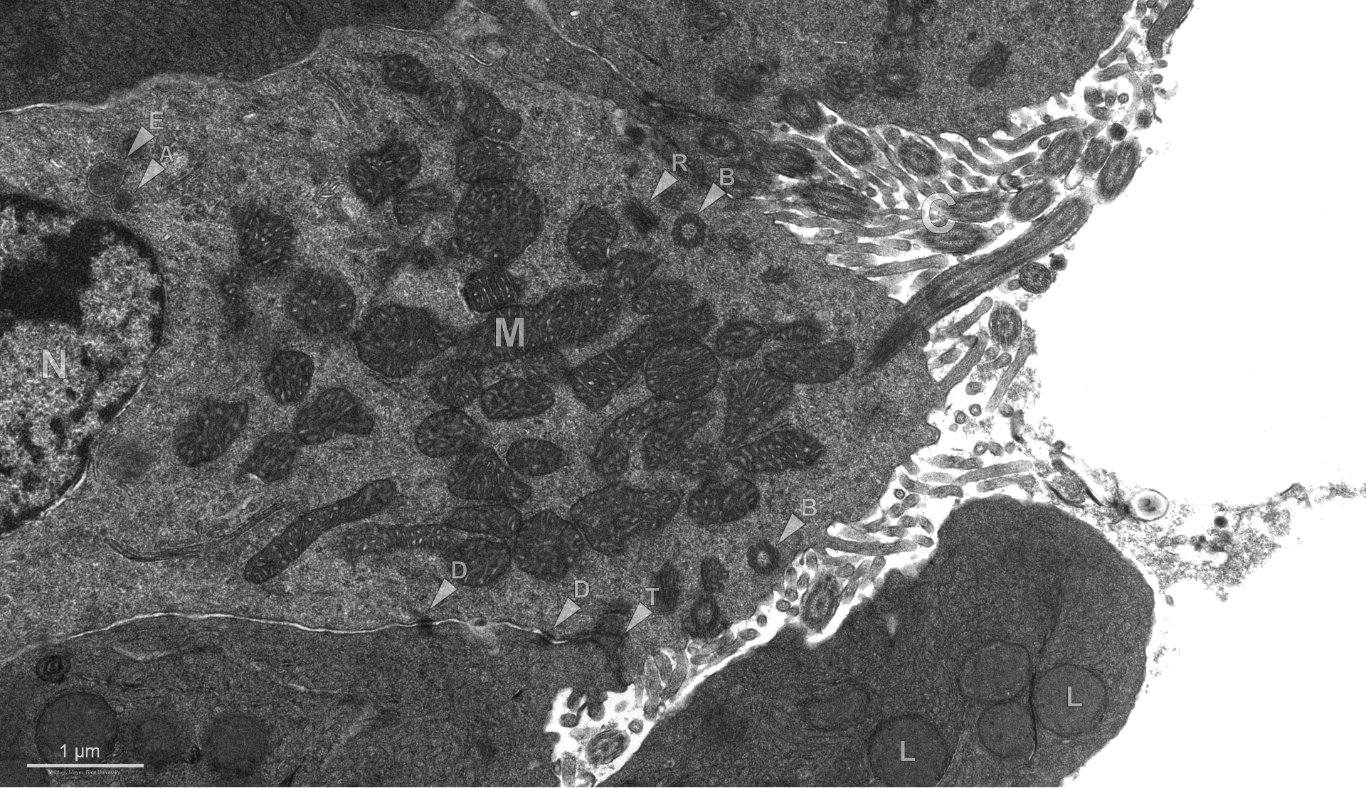
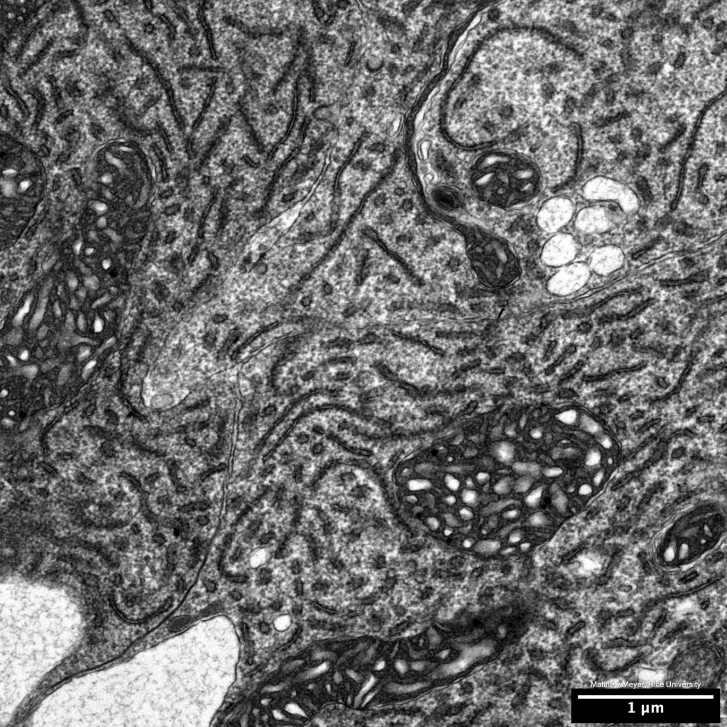
Lung epithelium. Nucleus (N), mitochondrial clustering (M), cilia (C), basal bodies (B), ciliary rootlet (R), desmosomes (D), tight junction complex (T), lipid droplets (L), autophagosomal membrane elongation nearing closure (E), autophagosome (A). Transmission electron microscopy (TEM) image.
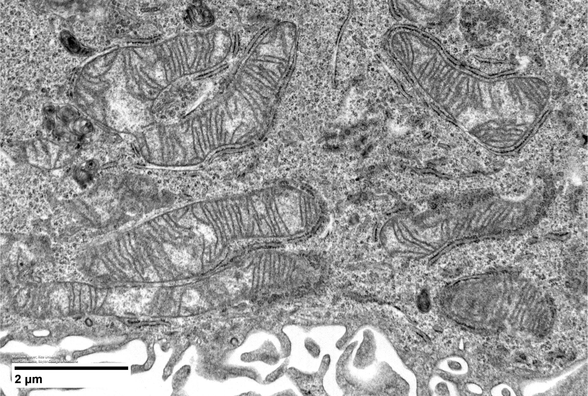
Cisplatin, a platinum-based chemotherapy drug, has been used to treat head and neck squamous cell carcinomas (HNSCC) and other solid tumors for over 30 years, despite a high frequency of drug resistance. In order to better treat HNSCCs it’s necessary to understand how this resistance works. In the lab, we found that cisplatin-resistant cells have reduced energy production. Interestingly, no alterations in mitochondrial integrity, morphology or number were detected in cisplatin-resistant cells! There is so much more to this story, including the role of KEAP1 mutations and RNA reductions with Nrf2 activation, so please check our our paper out now in the British Journal of Cancer (https://nature.com/articles/s41416-023-02253-7). Pictured above are mitochondria from HNSCC cell lines. It was an honor working with Vlad Sandulache and this massie collaborative team of scientists and physicians from @BCMFromtheLabs, @MDAndersonNews, @tamhsc, @UPMCHillmanCC & @Cornell!

Remember tardigrades, the microorganisms that can survive in outer space? They have a protein, Dsup, which protects their DNA from x-rays and harsh environmental stressors. Maybe humans can exploit Dsup to protect our neurons from neurodegenerative diseases? Here, we unfortunately discovered that Dsup promotes neurotoxicity and neurodegeneration in cortical neurons. Using electron microscopy, we discovered that Dsup promotes chromatin condensation. It was a pleasure working with Andrey Tsvetkov, Rocio Escarcega and the entire Tsvetkov lab on this paper! https://t.co/X9RWDajkb7
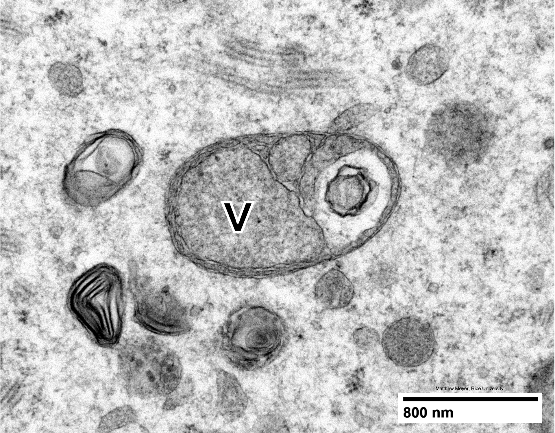
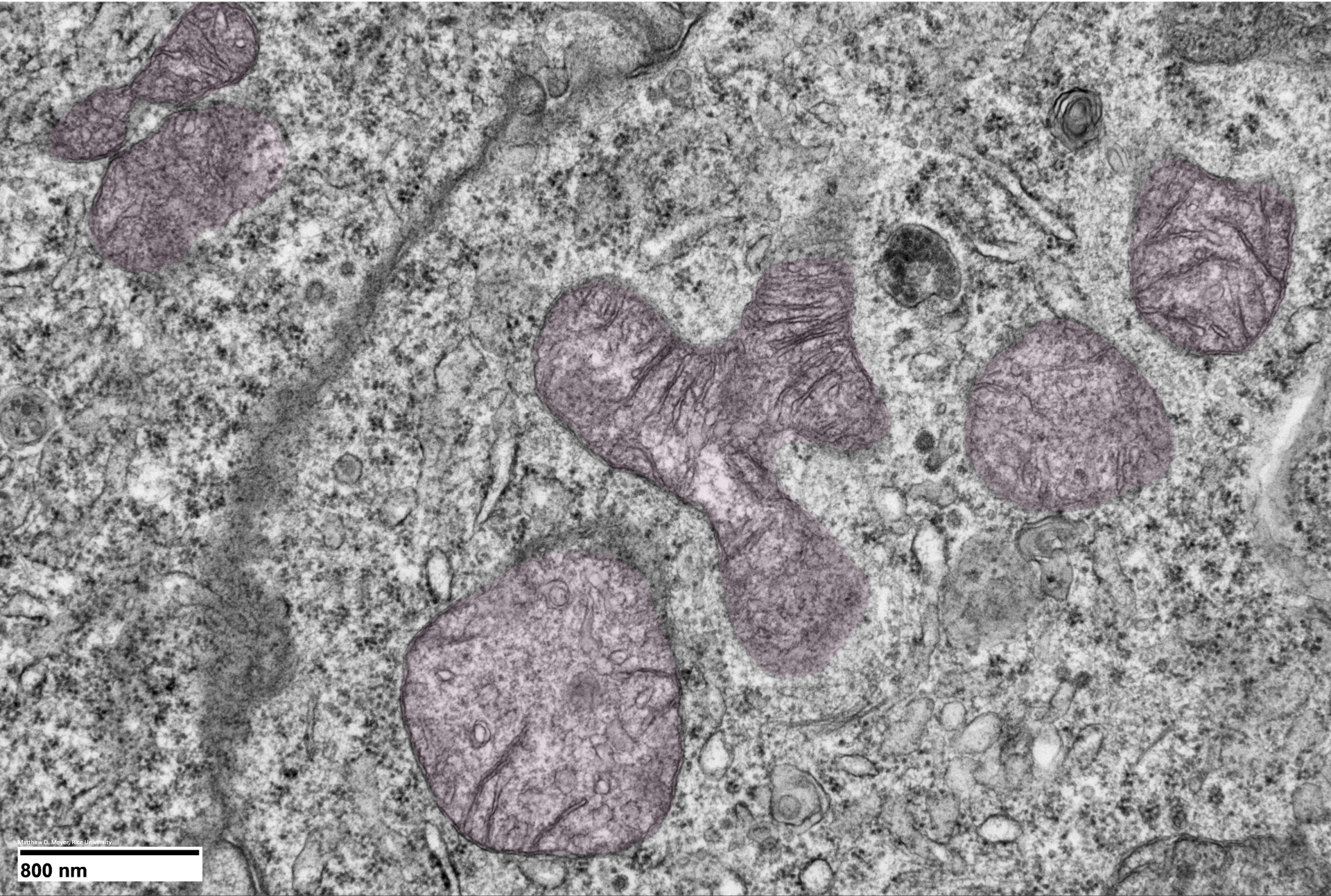
Mitochondria in oocytes are often morphologically unique, like this vacuolated (v) example.
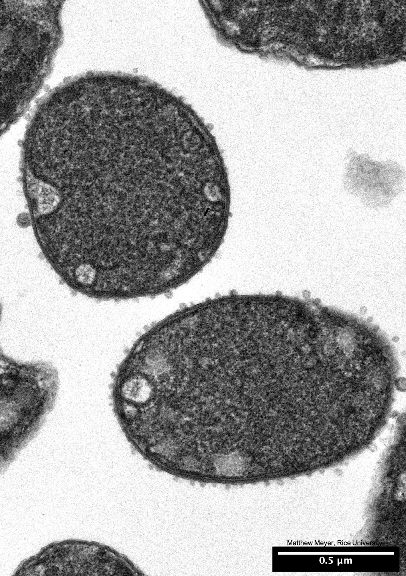
You probably have E. coli living in your gut. Here’s what this bacteria looks like when it’s sliced like a salami and thrown in a transmission electron microscope (TEM).
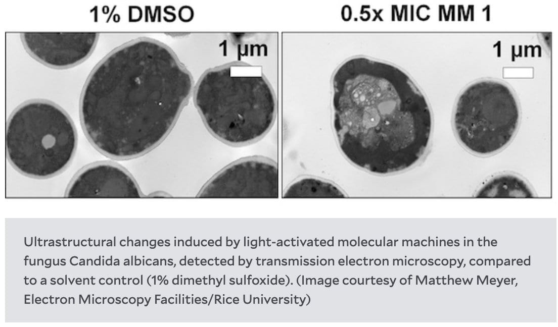
Molecular machines kill fungi by necrosis following mitochondrial dysfunction and calcium overload. Happy to make the TEM & SEM contribution to this paper! https://t.co/F3ev7dIsb1
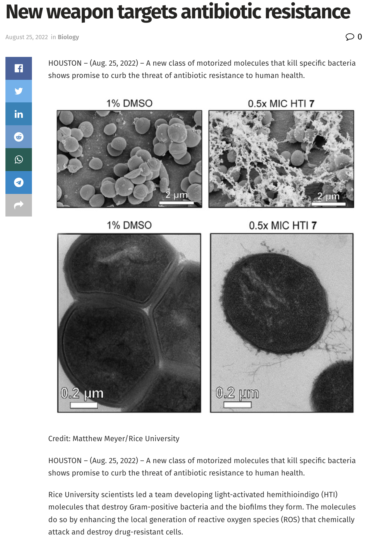
From: https://scienmag.com/new-weapon-targets-antibiotic-resistance/
TEM of mitochondria in breast cancer xenograft. Triple negative breastcancer that is resistant to chemotherapy may become more treatable if adaptations by mitochondria are overcame. It was a pleasure working with @DrGEcheverria and her extraordinary team on this project!
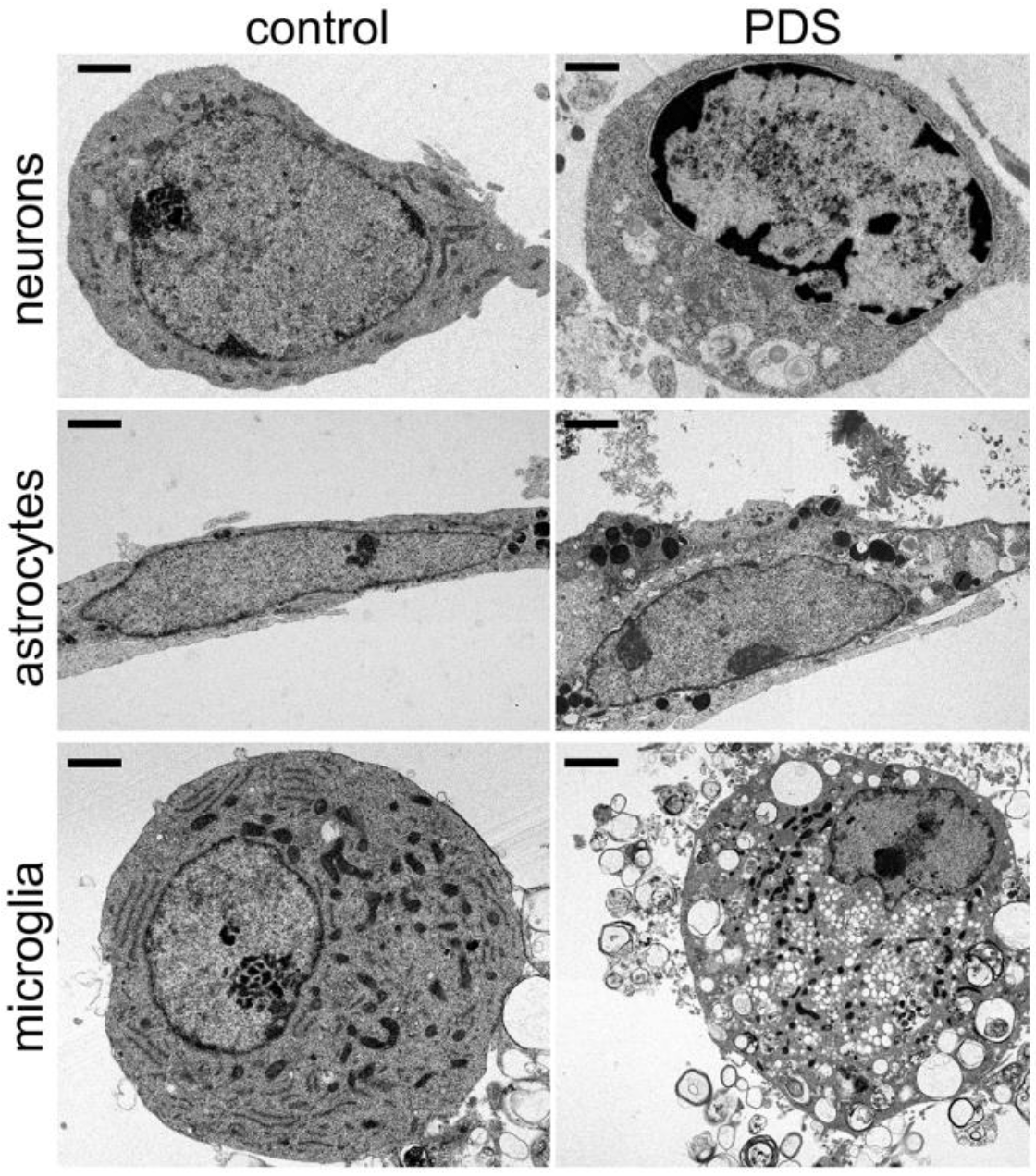
G4 DNA, when stabilized, alters chromatin and cytoplastic structure in neurons, astrocytes and microglia. This may help uncover a novel senescence pathway in brain aging. Happy to share our work with the Tsvetkov lab @McGovernMedin @AgingJrnl. https://t.co/KhjF9efyvK
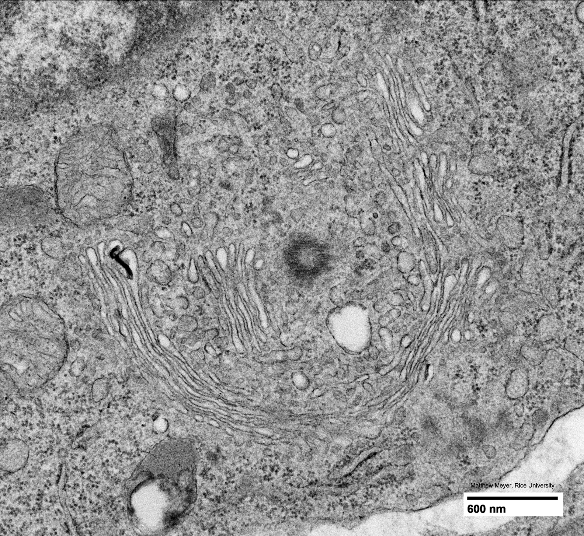
Golgi and Centriole
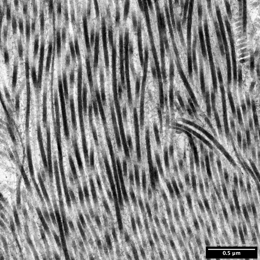
- Collagen is the most abundant protein in mammals. Here’s what it looks like in tissue, organized in collagen fibrils, which are semi-crystalline aggregates of collagen molecules.
- SEM image to two stem cells, in vitro.
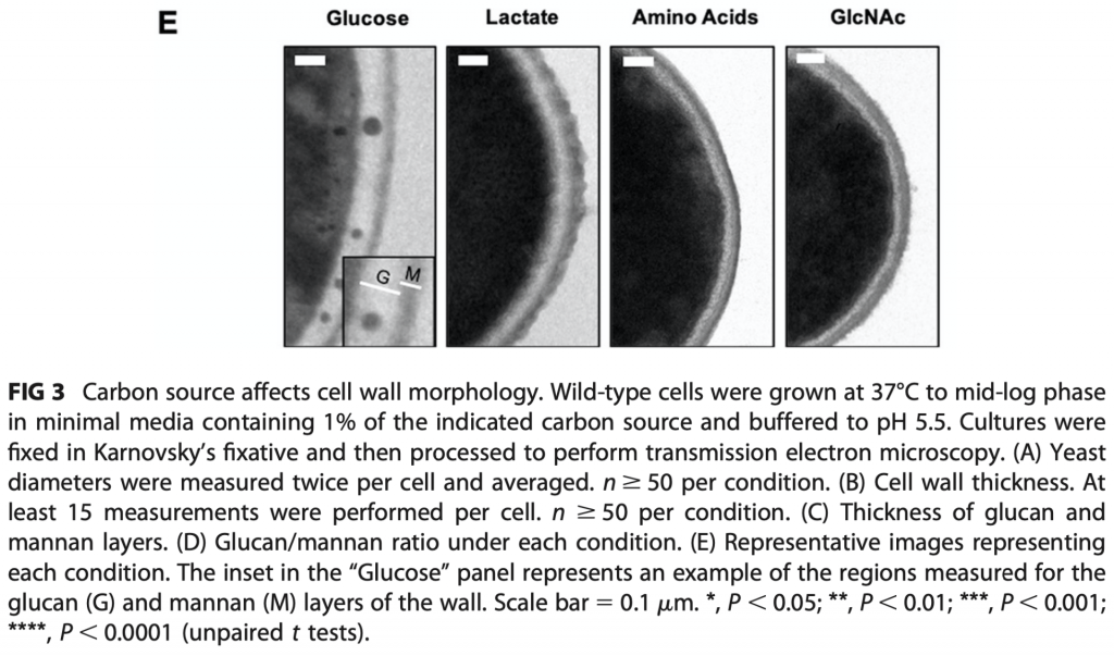
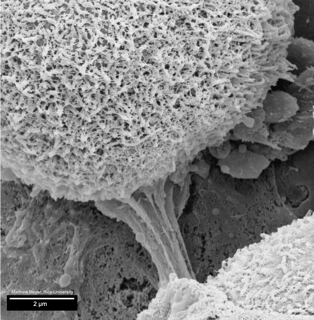
- Here, we provided TEM for Williams & Lorenz (DOI: 10.1128/mBio.03070-19), where they report that carbon source affects cell wall morphology in Candida albicans.

- We provided TEM for Jung et al. (DOI: 10.1002/adma.201908291), where they report that a multifunctional bio-nanocomposite coating for perishable fruits can be made from cellulose nanocrystal reinforced poly(albumen). These images show that coating.

- Stinging trichomes of a dewberry leaf, complete with trichome surface bacteria (outlined with white box).
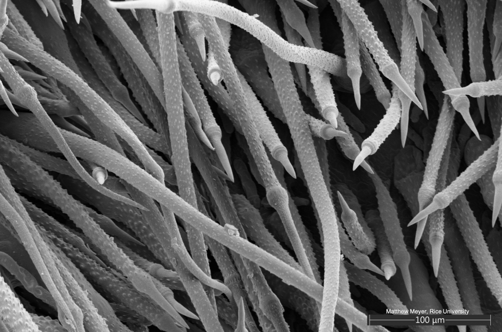
- Stinging trichomes of a dewberry leaf
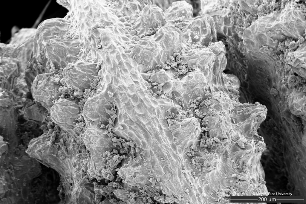
- Dewberry, Rubus aboriginum, collected at Rice University, outside the Space Science & Technology building. SEM images from Rice SEA’s new Thermo/FEI Apreo.
- Nissl bodies (and mitochondria) in zebrafish larval neurons.
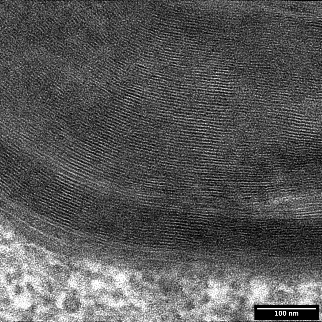
- Myelin
- Myelin
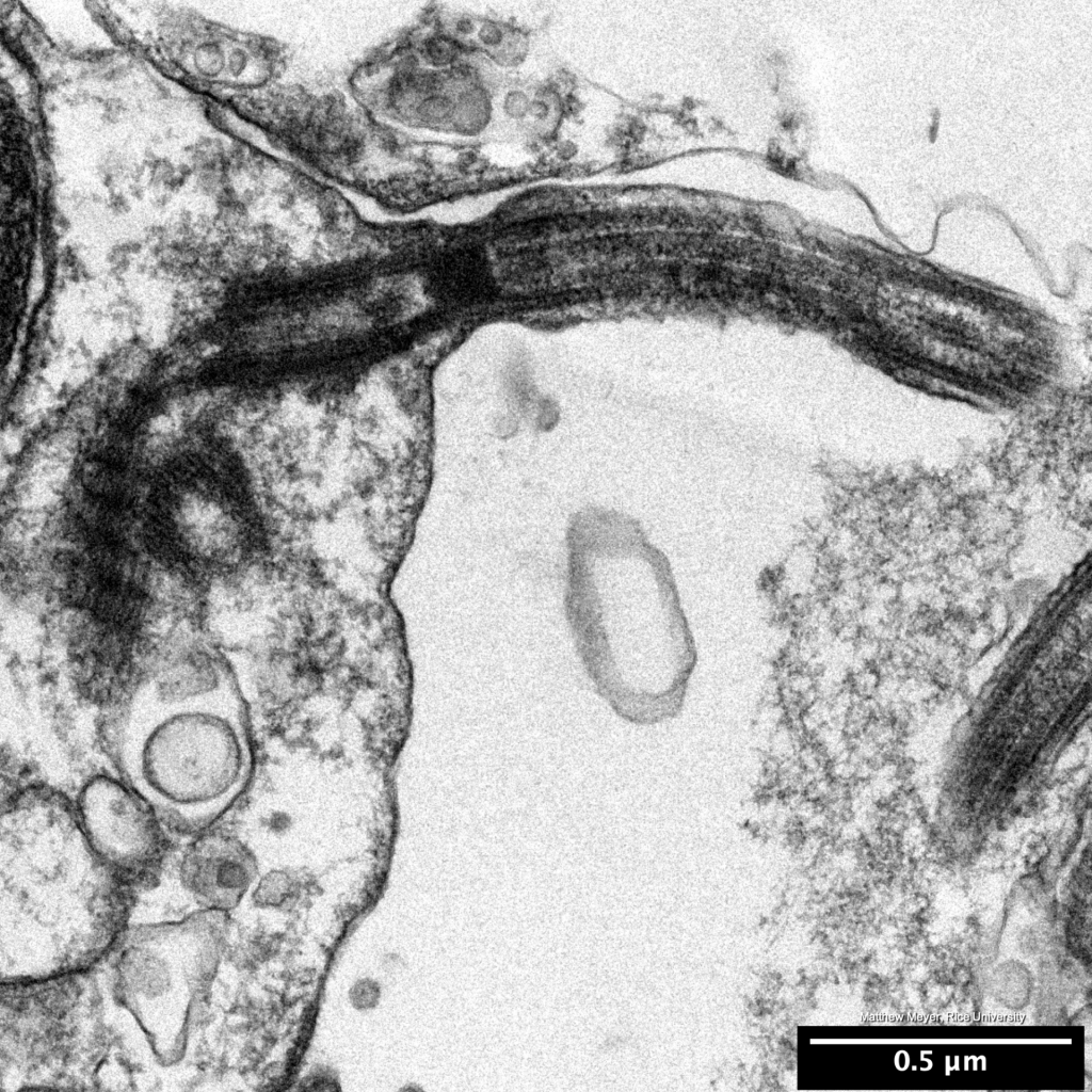
- Cilium, a TEM classic.
- Synaptic vesicles in adult zebrafish

- Synaptic vesicles in adult zebrafish

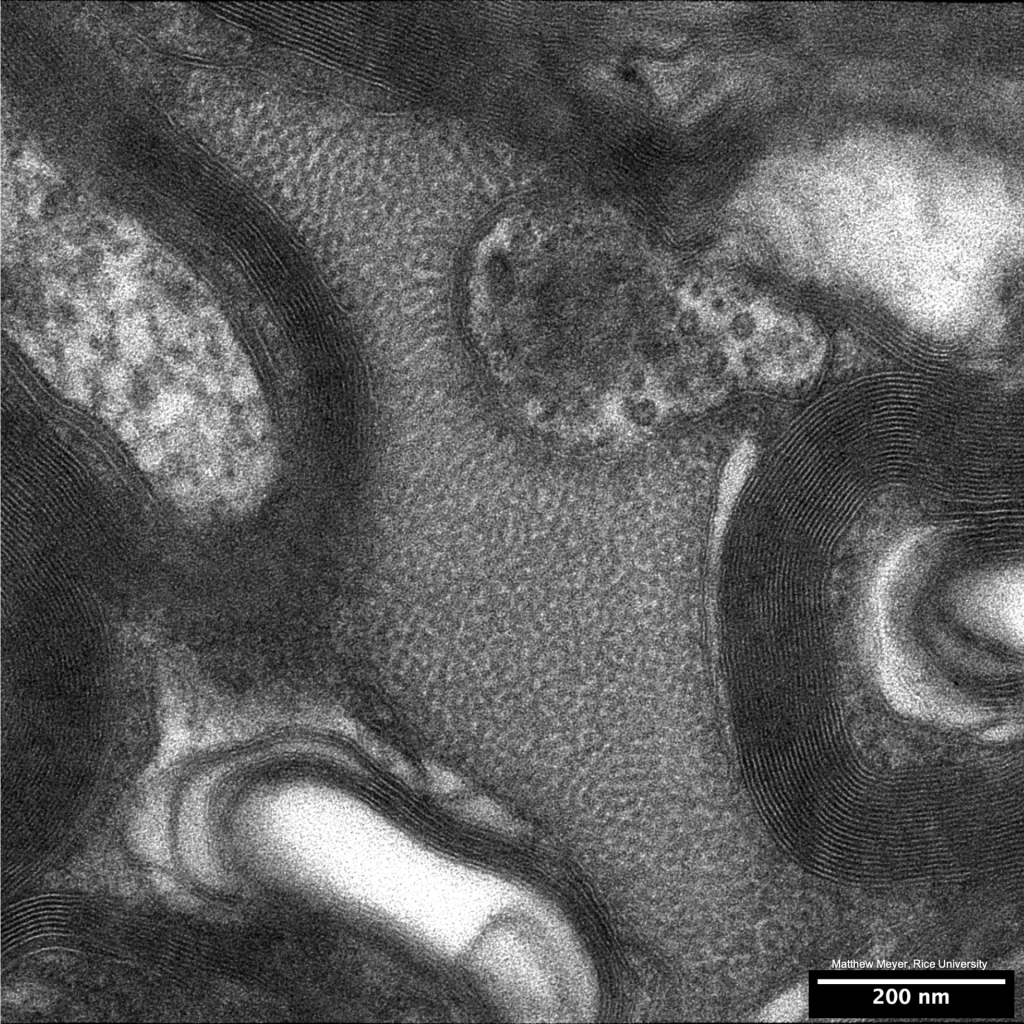
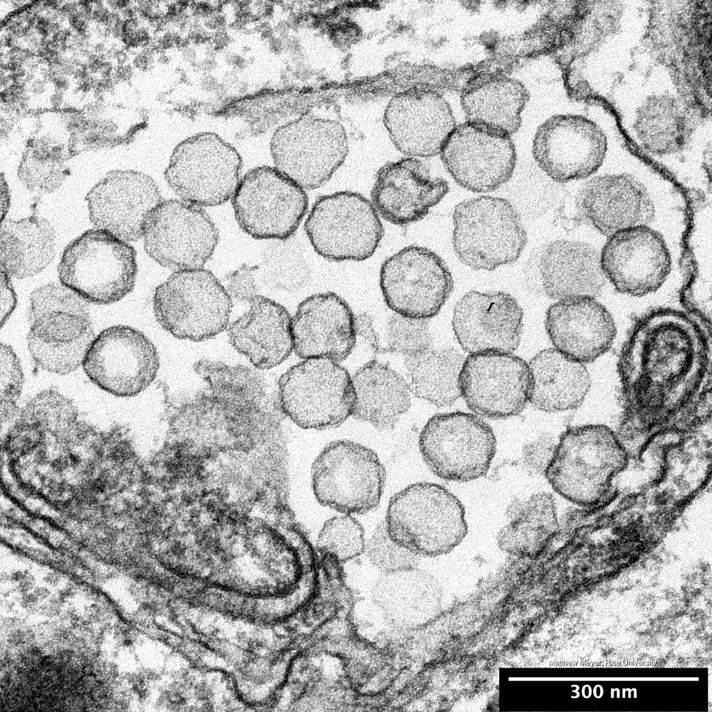
- Urate oxidase crystalline core of a peroxisome
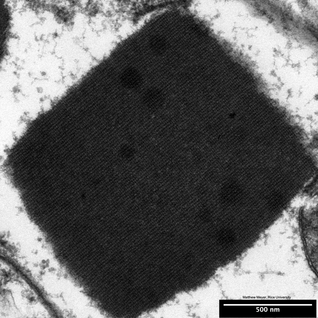
- Urate oxidase crystalline core of a peroxisome
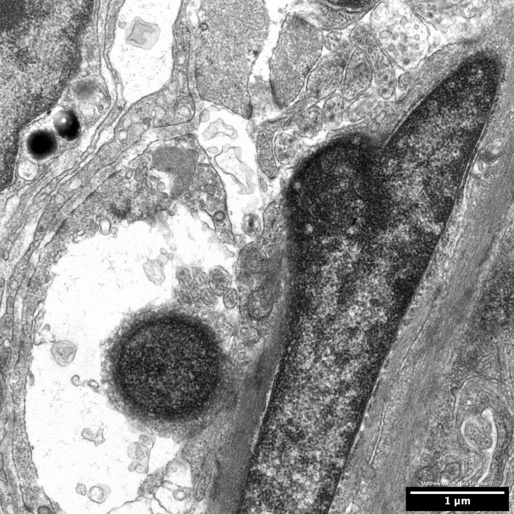
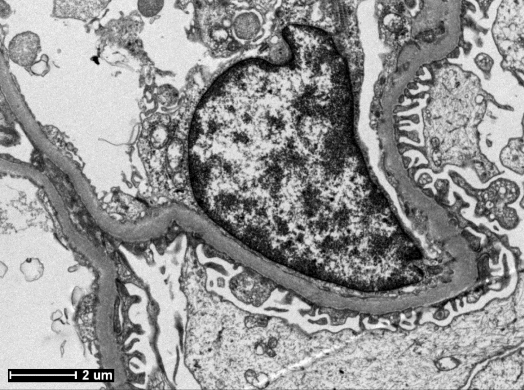
- An example of suspected nuclear elimination in zebrafish larvae
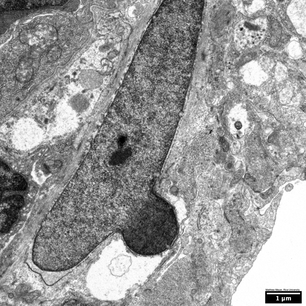
- An example of suspected nuclear elimination in zebrafish larvae
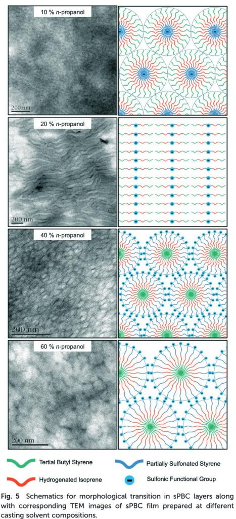
- We also take on electron microscopy projects involving non-biological samples. In this case: Self assembled, sulfonated pentablock copolymer cation exchange coatings for membrane capacitive deionization.
- TEM image of gut bacteria. Bacterial species unknown.

- TEM image of gut bacteria. Bacterial species unknown.
- An ionocyte in zebrafish larval epidermis
- Rodlet cell in zebrafish larval epidermis. Rodlet cell function is poorly understood.
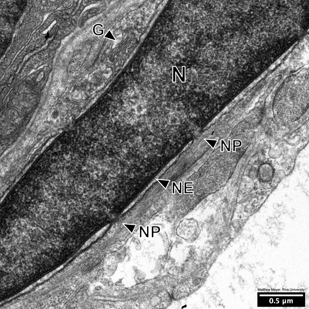
- A nucleus (N), with nice examples of nuclear pores (NP), along with the nuclear envelope (NE) and a Golgi apparatus (G).
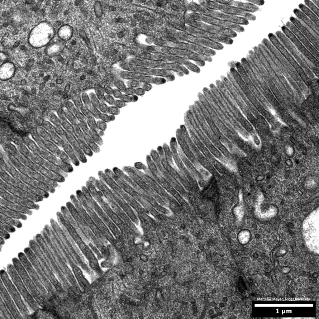
- Microvilli of the zebrafish gut
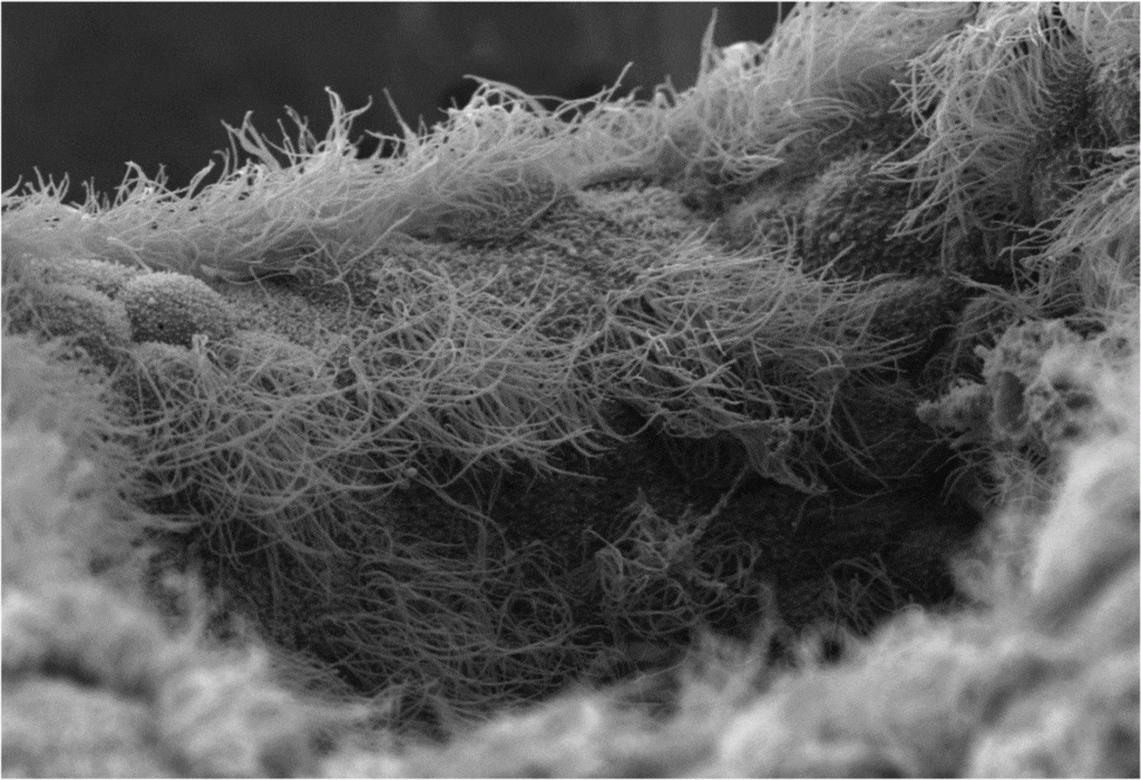
- Multiciliated cells of the developing nostril in Xenopus laevis larvae. SEM gif. Taken during my time in John Wallingford’s lab UT-Austin/HHMI
- Diagnostic Pathologists: Do your electron microscopy technicians need training? We provide training for renal bx adequacy, tissue processing, staining and TEM imaging. Matt is the former manager of clinical diagnostic TEM at the USC Keck School of Medicine.